42 onion cells under microscope with labels
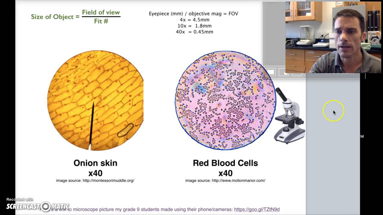
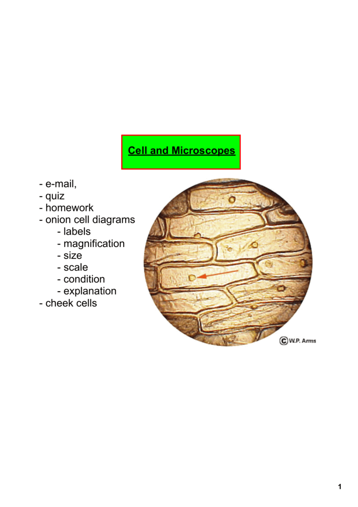
Onion Cells Under a Microscope - Requirements/Preparation/Observation Add a drop of iodine solution on the onion membrane (or methylene blue) Gently lay a microscopic cover slip on the membrane and press it down gently using a needle to remove air bubbles. Touch a blotting paper on one side of the slide to drain excess iodine/water solution, Place the slide on the microscope stage under low power to observe. PDF Onion Cells - Investigation - Exploring Nature 5. Observe the onion tissue under the microscope at 4x, 10x and 40x with lots of light (open diaphragm). Then slowly close the diaphragm while observing the image to find the best light for seeing cellular details. 6. Draw a section of onion skin cells at 10x magnification. Then switch to 40x and draw one cell and label it. Questions: 1.
Onion Root Tip Mitosis - Stages, Experiment and Results · Cover the sample (root tip) with a coverslip and gently press the coverslip down, then examine the slide under the microscope starting with low magnification * For this experiment, a properly prepared slide should appear light pink due to the stain to almost colorless. * Unused roots can be stored in 70 percent alcohol. Results

Onion cells under microscope with labels
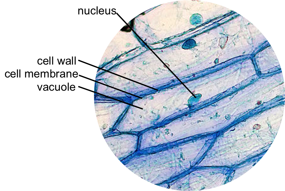
Natural Sciences Grade 9 - Grade 7-9 Workbooks The onion cells have a thick cell wall and a cell membrane. The animal cells only have a cell membrane. The onion cells have a regular shape whereas the cheek cells have a irregular shape and seem more flimsy. In the onion cells they might notice a large vacuole which might not be as visible in the cheek cells. Cheek cells do not have vacuoles ... Onion Epidermis - kuensting.org Onion epidermal cells, iodine stain, 400X. The nucleus of an onion epidermal cell, 1000X magnification. ... Microscopy, size and magnification - Microscopy, size and ... - BBC Place cells on a microscope slide. Add a drop of water or iodine (a chemical stain). Lower a coverslip onto the onion cells using forceps or a mounted needle. This needs to be done gently to...
Onion cells under microscope with labels. Health & Safety Meeting Dates | Institute Of Infectious ... Feb 08, 2022 · IDM H&S committee meetings for 2022 will be held via Microsoft Teams on the following Tuesdays at 12h30-13h30: 8 February 2022; 31 May 2022; 2 August 2022 The Cell - ScienceQuiz.net The diagram shows a group of onion cells. The parts labelled A, B and C respectively are ... The diagram shows a plant cell as seen under a microscope. Two of the ... Cells and Reproduction - BBC Bitesize Onion cells are easy to see using a light microscope. ... A small tube placed under the skin of the upper arm. ... Five small tubes with labels and stoppers or lids Cress seeds Labels Cotton wool ... Epidermal onion cells under a microscope. Plant cells appear polygonal ... Observing onion cells under the microscope. For this microscope experiment, the thin membrane will be used to observe the cells. An easy beginner experiment. Jessica Williams. Ideas for Work. Similar ideas popular now. Applied Science. Subjects. Physical Science. Technology.
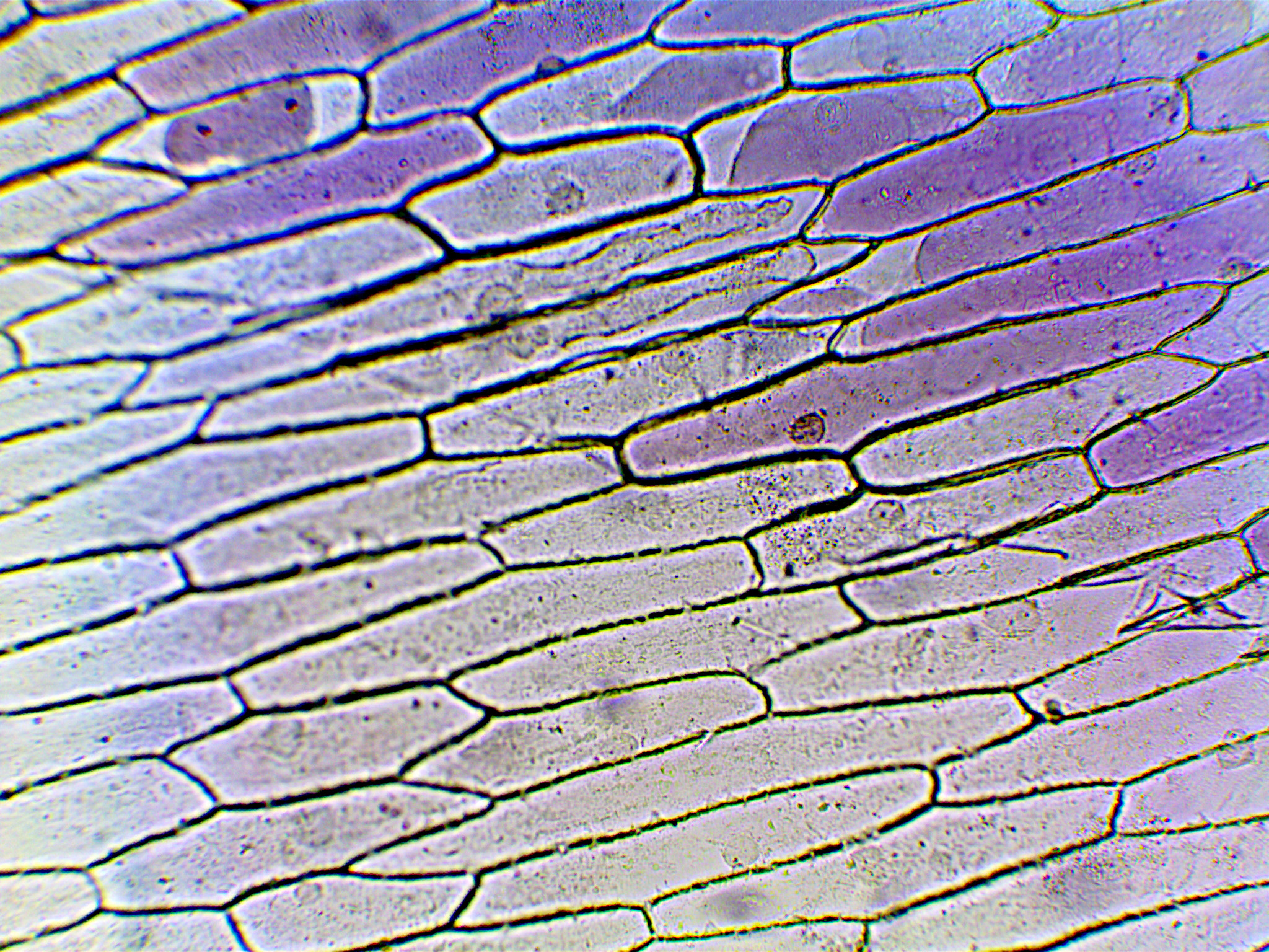
Onion Skin Cells Labeled - the wonderful microworld onion skin cells ... Onion Skin Cells Labeled. Here are a number of highest rated Onion Skin Cells Labeled pictures on internet. We identified it from obedient source. Its submitted by executive in the best field. We... Oxford Cambridge and RSA Friday 16 October 2020 – Morning 1 (a) A student was observing onion epithelial cells using a light microscope. They photographed these cells and the image obtained is shown in Fig. 1.1. The student then made a drawing of a few cells from this image. The drawing is shown in Fig. 1.2. Fig. 1.1 cytoplasm cell wall large permanent vacuole ribosome Fig. 1.2 Onion Cell Lab Report.docx - Onion Cell Lab Report By Onion Cell Lab Report By : Nawaf Almalki Introduction: Many things that are viewed using a microscope, particularly cells, can appear quite transparent under the microscope. The internal parts of the cells, the organelles, are so transparent that they are often difficult to see. Biologists have developed a number of stains that help them see the cells and their organelles by adding color to ... Onion Microscope Under Cell Labeled Search: Onion Cell Under Microscope Labeled. Add 2 drops of iodine (or other stain) to the onion slide Use the microscope, slide of "JF", mm ruler and photo of onion cells to assist you in answering the questions You will first view the cell under normal conditions, so you can easily be compared to the results if a change occurs Sketch of one cell in each phase of mitosis (prophase, metaphase ...
Observing Onion Cells Under The Microscope Afterwards, carefully mount the prepared and stained onion cell slide onto the microscope stage. Make sure that the cover slip is perfectly aligned with the microscope slide, and that any excess stain has been wiped off. Secure the slide on the stage using the stage clips. My onion cells at 40x magnification :) | Cellular level, Abstract, Neon ... This low magnification image shows an insular/organoid growth pattern and extensive areas of necrosis. Any monkey can run an equation through a graphing calculator, call it "Icosahedron 12" and sell a "giclee image" for $300 to a gullible sophomore. But it takes actual smarts (or a serious bronze foundry) to make cool "science art." Ramin, 13 ... Looking at the Structure of Cells in the Microscope Both types of light microscopy are widely used to visualize living cells. Figure 9-7 Two ways to obtain contrast in light microscopy. (A) The stained portions of the cell reduce the amplitude of light waves of particular wavelengths passing through them. A colored image of the cell is thereby obtained that is visible in the ordinary way. (more...) Lennox Educational 01: To use a light microscope; 02: To obtain a good specimen of plant tissue for viewing under the microscope (onion cells) 03: To obtain a good specimen of animal tissue for viewing under the microscope (cheek cells) 04: To investigate the digestion of starch by amylase; 05: To investigate the effect of exercise on heart rate
SENTHIL PRABHU SIVASAMY: Observation of Plant & Animal Cells
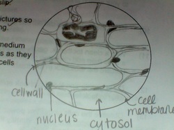
DOC Plant and Animal Cells Microscope Lab - Hillsboro City Schools Make a drawing of one onion cell, labeling all of its parts as you observe them. (At minimum you should observe the nucleus, cell wall, and cytoplasm.) Cheek cells 1. To view cheek cells, gently scrape the inside lining of your cheek with a toothpick. DO NOT GOUGE THE INSIDE OF YOUR CHEEK! (We will observe blood cells in a future lab!!) 2.

Living and Learning: Testing out my Microscope
onion cells under a microscope labeled - cahierdeseoul.com RM BRC1AJ - Light Micrograph (LM) of onion skin cells, magnification x 600. Pay close attention, you'll need to label cell slides on the test. Clean the stain from the slide and cover glass. Observe the onion tissue under the microscope at 4x, 10x and 40x with lots of light (open diaphragm). 7. 2. Onion cells under the microscope.

Phases of mitosis in control and 1-day CA-treated root tip cells. Bars... | Download Scientific ...
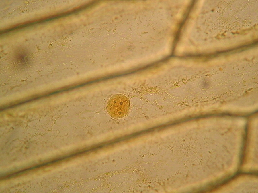
DOC The Onion Cell Lab - chsd.us Onion tissue provides excellent cells to study under the microscope. The main cell structures are easy to see when viewed with the microscope at medium power. For example, you will observe a large circular . nucleus. in each cell, which contains the genetic material for the cell. In each nucleus, are round bodies called . nucleoli

onion cells through microscope | I put my camera right up to… | Flickr
Onion Under Microscope Labeled Cell A drawing of one of the cells as seen under high power is shown below Observing Cells Under Microscope Obtain a slide of onion root cells Obtain a slide of onion root cells. Label any and all parts you can identify (512) 585-1153 (cell) Kayak Committee Fred Wahlers cell 214-476-7725 [email protected] Chemicals for microscopy include buffers ...

swifty science: onion cell lab
The Biology Project The Biology Project, an interactive online resource for learning biology developed at The University of Arizona. The Biology Project is fun, richly illustrated, and tested on 1000s of students.

Rens blog : Science, cells
PDF Onion Cell Lab - somewaresinmaine.com Research Biology Onion Cell Lab page 1 of 3 Onion Cell Lab After you have completed the rest of this lab come back to this cover page DRAW & LABEL AN ONION CELL WITH ALL THE PARTS / ORGANELLES YOU OBSERVE UNDER 40X. Purpose: To observe and identify major plant cell structures and to relate the structure of the cell to its function. Materials: 1 ...
Onion Cell Under Microscope 4x 10x 40x - Micropedia
The following diagram shows cells of onion peel label class ... - Vedantu 115.2k + views. Hint: The diagrams mentioned above are the internal structure of an onion peel and human cheek cells. In order to label them, we need to understand its anatomy and know about various structures present in it. Onion peel is an example of a plant cell whereas a human cheek cell is an example of an animal cell. Complete answer:

Onion Cell Under Microscope - Personal Experience with Microscopes - AyushiSinhaMicroscopy ...
Under the Micrsocope: Onion Cell (100x - 400x) - YouTube In this "experiment" we will see onion cells under the microscope.For the experiment you will only need onion, dropper and the microscope (container and tool...
The inner epidermis of the onion bulb’s cataphylls (the onion skin).
Plant tissue under a microscope - xylem and phloem - Rs' Science The highly active mitosis area is highlighted with a red dash line. Within that area, you can easily find cells undergoing different phases of mitosis, prophase , metaphase , anaphase, and telophase. (Modified from the guidebook of Rs' Science - 25 Microscope Prepared Slide Set) The Stem - Xylem and Phloem

The Cells and Microorganisms Webquest
Onion cell Images, Stock Photos & Vectors - Shutterstock Find Onion cell stock images in HD and millions of other royalty-free stock photos, illustrations and vectors in the Shutterstock collection. Thousands of new, high-quality pictures added every day.
The Cell — The Biology Primer
Onion Skin Cells Labeled - chapter 7, the wonderful microworld onion ... Onion Skin Cells Labeled. Here are a number of highest rated Onion Skin Cells Labeled pictures on internet. We identified it from reliable source. Its submitted by presidency in the best field. We...
Onion Cells under Microscope
What organelles are in an onion cell? - Biology Stack Exchange To answer your question, onion cells (you usually use epithelial cells for this experiment) are 'normal' cells with all of the 'normal' organelles: nucleus, cytoplasm, cell wall and membrane, mitochondria, ribosomes, rough and smooth endoplasmic reticulum, centrioles, Golgi body and vacuoles.
Labeled Onion Cell Under Microscope 40x - Micropedia
Plant Cell Under Microscope Labeled 40X : Young Root 2 Of Broad Bean ... Cells and viewing them under the microscope. A small square of a red onion skin (membrane) was observed under a microscope at high power (x40) magnification. (iv) describe how you applied the stain. They must draw and label the nucleus, cell membrane set up your microscope, place the onion root slide on the stage and focus on low (40x) power.
Microscope Onion Cell Labeled - Micropedia
Onion Cells Under a Microscope (100x-2500x) - YouTube In this video you will see onion cells under a microscope (100x-2500x) as is, without any coloring. To observe the onion cells the thin membrane is used. It...
Post a Comment for "42 onion cells under microscope with labels"